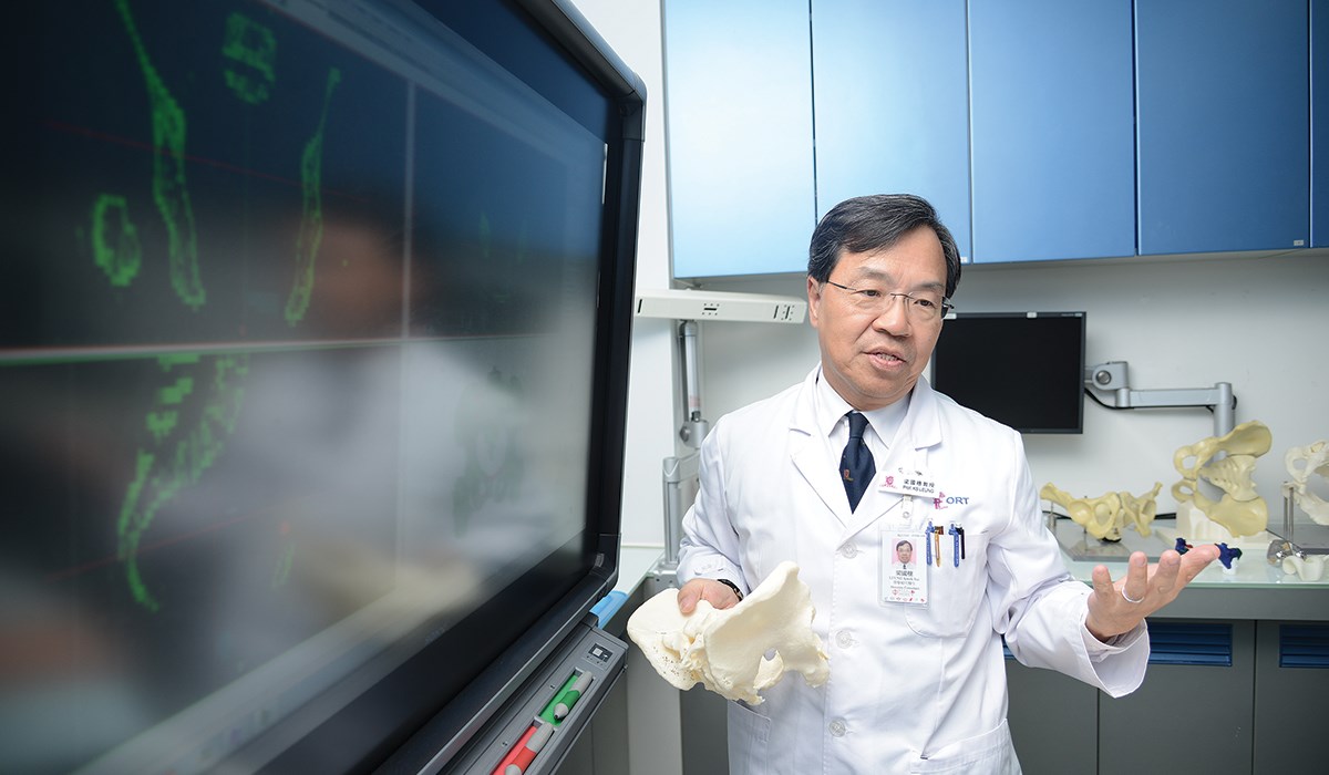
Professor Kwok-sui Leung is expanding his department of Orthopaedics and Traumatology, Faculty of Medicine of the Chinese University of Hong Kong (CUHK) beyond its traditional role as an academic clinical department. At the department’s Prince of Wales Hospital, Leung is improving the success and accuracy of surgeries, boosting patient confidence and exploring new innovative surgical methods all at once. How? By using 3D printing to create bone models and surgical guides for orthopedic surgery preparation.
The Prince of Wales Hospital adopted a Fortus® 3D Production System that uses FDM Technology™ to print both surgical guides and surgical tools. Before using 3D models to prepare for surgical procedures, most surgeons at the Prince of Wales Hospital relied on their experience and CT scans to visualize and plan the operation. Validating their planned approach took place in the operating room. Preparing for an operation using 3D printed models shortens the surgical process and increases operation accuracy and success rate.
One of the most common incision sites in bone cancer surgeries is at the end of the femur, close to the knee joint. Accuracy of an incision site is critical to completely remove the tumor and reconstruct a functioning extremity. Correcting pelvic fractures from car accidents – another routine hospital procedure – also requires extreme precision. For complex procedures, such as determining the angle of screw placement and location of metal implants, 3D models provide the trial space necessary to gain the accuracy desired.
Prince of Wales Hospital uses 3D printing in cases ranging from corrective osteotomy (re-alignment of bone from deformity) to complex bone fractures from car accidents. On average, operation time was reduced by an hour when incorporating 3D printed parts in the pre-surgical process.
Elvis Chun-sing Chui, biomedical engineer at the Orthopaedic Learning Centre of CUHK, said, “The adoption of 3D printing provides a platform to experiment with innovative surgical approaches. Moreover, it enhances the communication between medical practitioners and patients. Patients better understand the diagnosis and treatments with the aid of the 3D printed parts.”
In addition to printing surgical guides, CUHK and Prince of Wales Hospital have further extended their applications to printing small, single-use surgical tools.
“3D printing helps us advance medical research and development,” said Leung. “It [3D printing] offers ample potential in both the surgical guides and surgical tools areas. And, the cost saved from using 3D printing can now be used for research and development, ultimately benefiting patients, surgeons and researchers.”

 to complex bone fractures from car accidents, like the above.")


